State of the Art Dental Technology
Digital
X-Rays
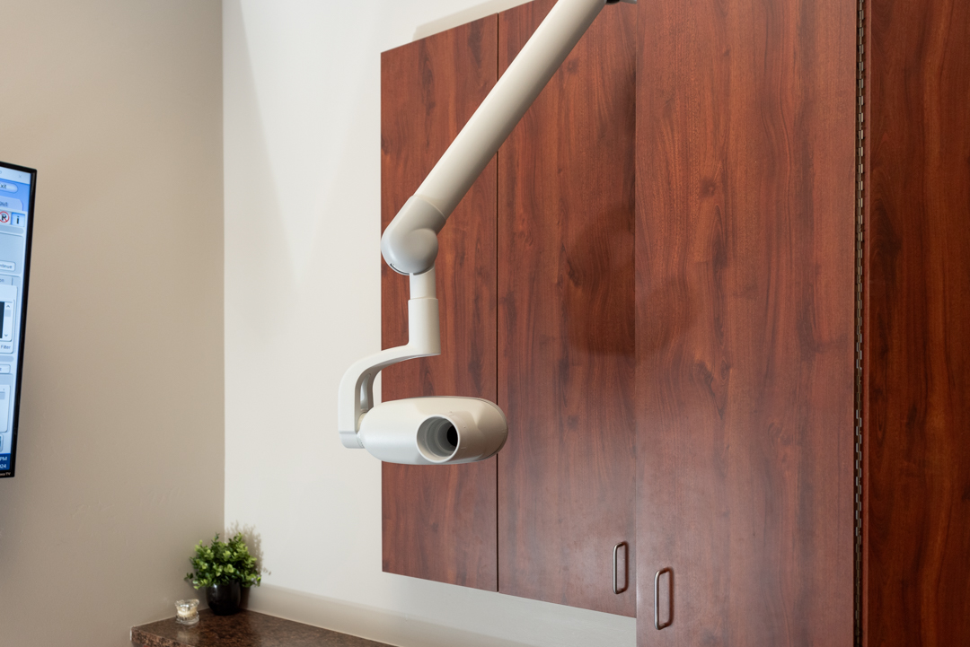
Digital x-rays use a digital image capture device in place of traditional film, sending an image immediately to a computer. The result is a highly-detailed image of the mouth, and its contrast and resolution can be enhanced to more easily diagnose dental problems and determine the best treatment with less radiation.

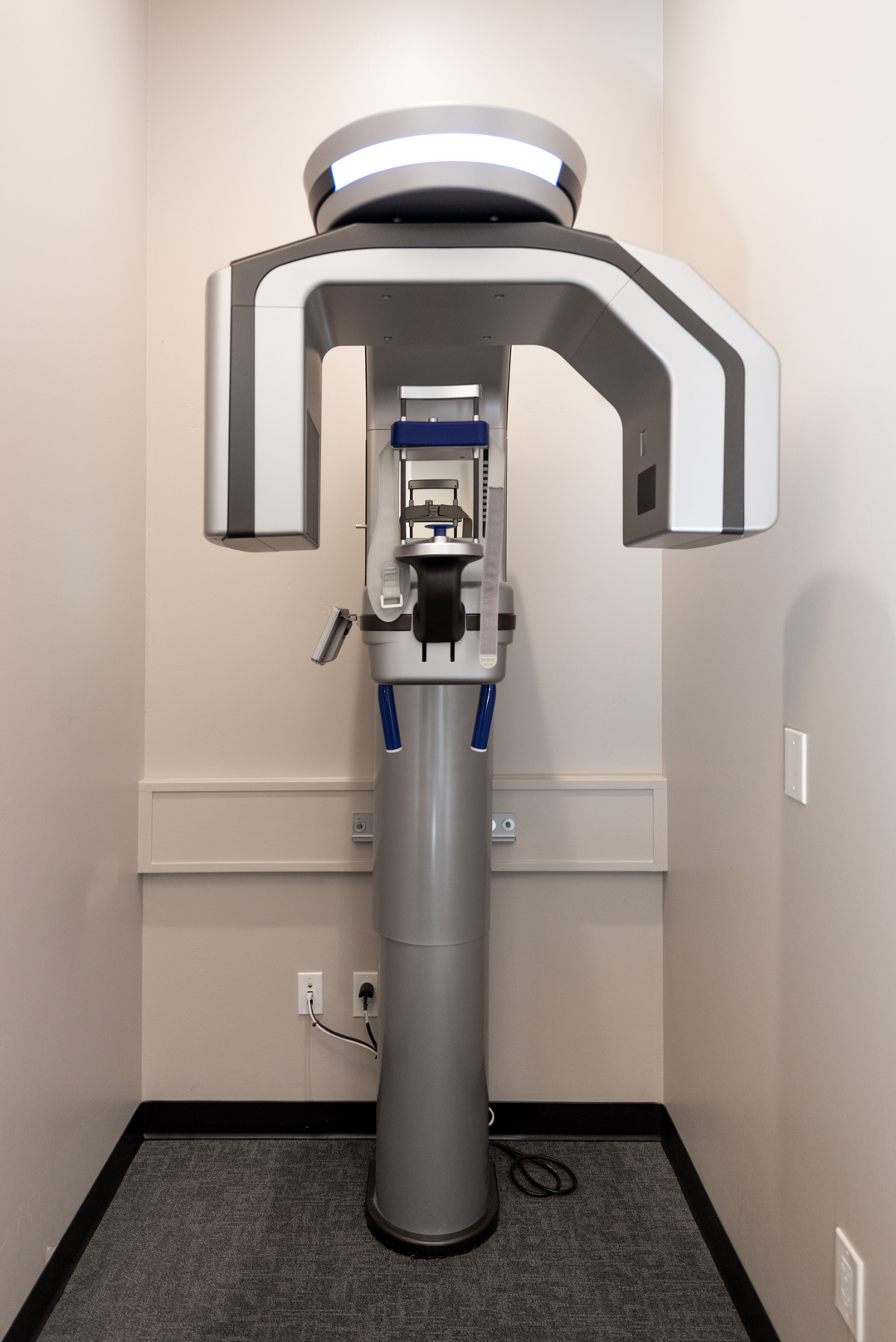
Prexion Excelsior CBCT
We use our CBCT to produce three dimensional (3-D) images of your teeth, soft tissues, nerve pathways and bone in a single scan that takes 12 seconds once the scan is started. It allows Dr. Phelps to see a detailed picture from many angles so he can properly diagnose what is going on in the patient's mouth. Up to 1/3 of abscesses can be missed on a 2D digital x-ray. With a CBCT the infection is more visible. The CBCT allows for comprehensive views of width and depth of bone levels needed for implant planning. What may have been a mystery can now be clearly seen and diagnosed leading to a speedy treatment and better long term prognosis.


